


For transparency and scientific grounding, we invite you to explore the published studies that support our work.
Our research and clinical insights are supported by peer-reviewed scientific studies and published literature. Below you can explore two categories of references:
These references provide the scientific foundation behind our work and reflect the continuous progress in ophthalmic research.
This study investigates the efficacy of Ultraviolet C (UV-C) light therapy as a potential treatment for bacterial keratitis. Conducted under controlled in vitro conditions, the research evaluates the antimicrobial activity and safety of UV-C exposure on affected corneal tissue. The findings contribute to the growing body of evidence supporting UV-C therapy as a non-invasive and targeted approach for managing severe corneal infections in veterinary ophthalmology.
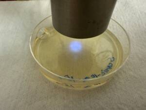
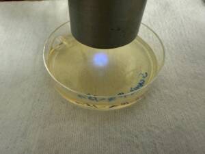
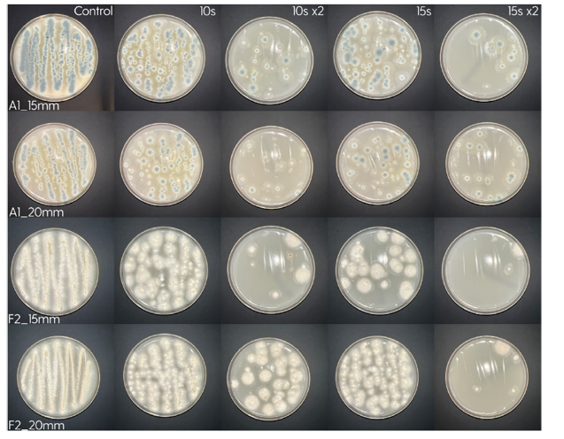
Treatment of the Petri plates inoculated with live bacteria using Ocu-Vet UV-C for 15 seconds at a 10 mm distance.
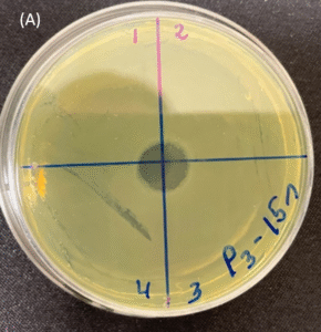
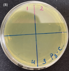
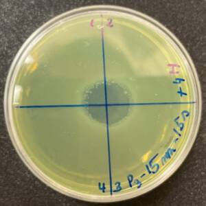
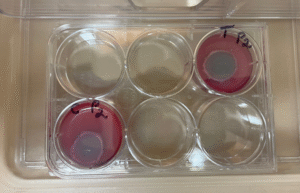
Photos of the Petri plates, 72 hours after bacterial (Pseudomonas aeruginosa) inoculation.
Image (A) shows positive bacterial growth inhibition (full translucency) at the treatment site after one dose of
UV-C for 15 seconds at 10 mm distance.
Image (B) is the control plate. The blue lines divide the plate into quadrants.
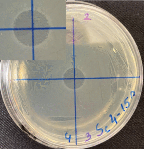
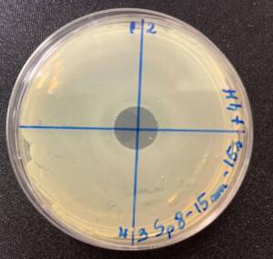
Photos of the Petri plates, 72 hours after bacterial (Streptococcus canis) inoculation.
The image and the inset show complete (positive) bacterial growth inhibition (full translucency)
at the treatment site after one dose of UV-C for 15 seconds at a 10 mm distance.
The blue lines divide the plate into quadrants.
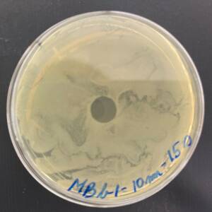
Photo of the Petri plate, 72 hours after bacterial (Moraxella bovis) inoculation. The image shows complete (positive) bacterial growth inhibition (full translucency) at the treatment site after one dose of UV-C for 15 seconds at a 10 mm distance.
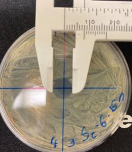
The diameter of the area of inhibition, measured with calipers:
– 8 mm (7 mm for Staphylococcus isolates) at 15 seconds.
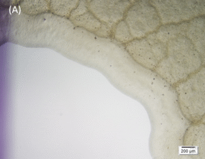
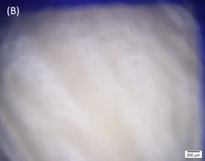
Microscopic images (4× objective, 5× magnification, Olympus cellSens Standard software) of the central area inoculated with Pseudomonas aeruginosa and treated with UV-C for 15 seconds (A) and the control area (B). A clear demarcation of the treated area can be seen on image (A).
Photos of the Petri plates, 72 hours after bacterial inoculation. Both plates show complete (positive) bacterial growth inhibition (full translucency) at the treatment site after two UV-C doses of 15 seconds at a 15 mm distance. The blue lines divide the plate into quadrants.
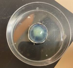
Mimicking a superficial ulcer by scoring and removing the epithelium and anterior stroma of the canine cornea. Fluorescein staining was used to confirm the presence of the superficial ulcer.
Canine cornea inoculated superficially on the ulcer bed with live Pseudomonas aeruginosa bacteria.
Storage of inoculated cornea and control in the incubator for 24 hours. The corneas are immersed in a storage solution to prevent desiccation. The image shows an inoculated cornea with live bacteria.
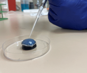
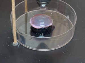
Treatment of the inoculated cornea with UV-C for 15 seconds at a distance of 10 mm.
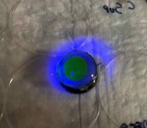
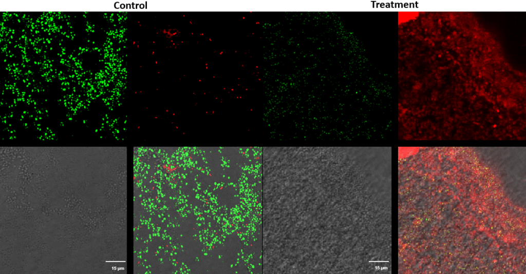
Staphylococcus pseudintermedius – Superficial fluorescence microscope images showing Staphylococcus pseudintermedius inoculated on the cornea, treated with UV-C for 15 seconds at a 10 mm distance.
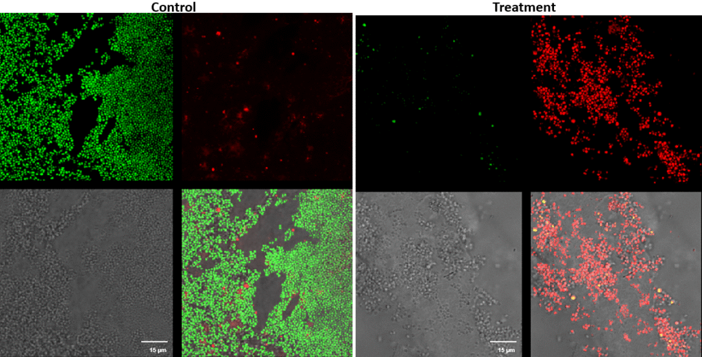
Streptococcus canis – Superficial fluorescence microscopy. Each image is divided into four quadrants: the upper left quadrant shows the green channel highlighting live bacteria; the upper right shows the red channel highlighting dead bacteria; the bottom right shows the merged green and red channels indicating live and dead bacteria; and the bottom left illustrates the bright field image. Bright green represents bacteria with intact membranes (live), red represents bacteria with compromised or dead membranes, and yellow represents bacteria with partially compromised membranes. Scale bar: 15 µm.
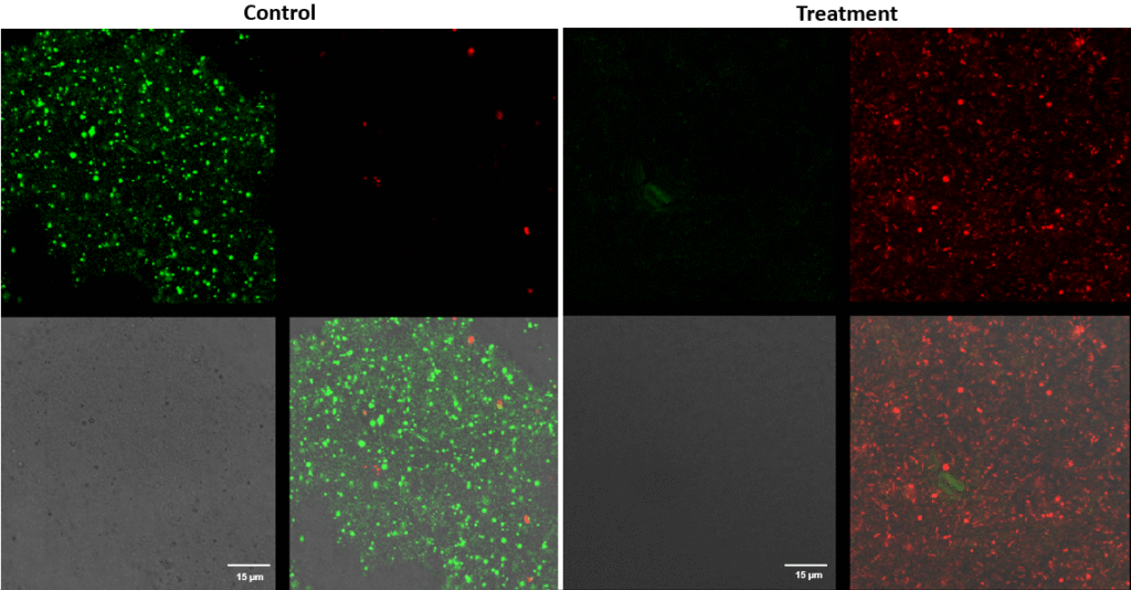
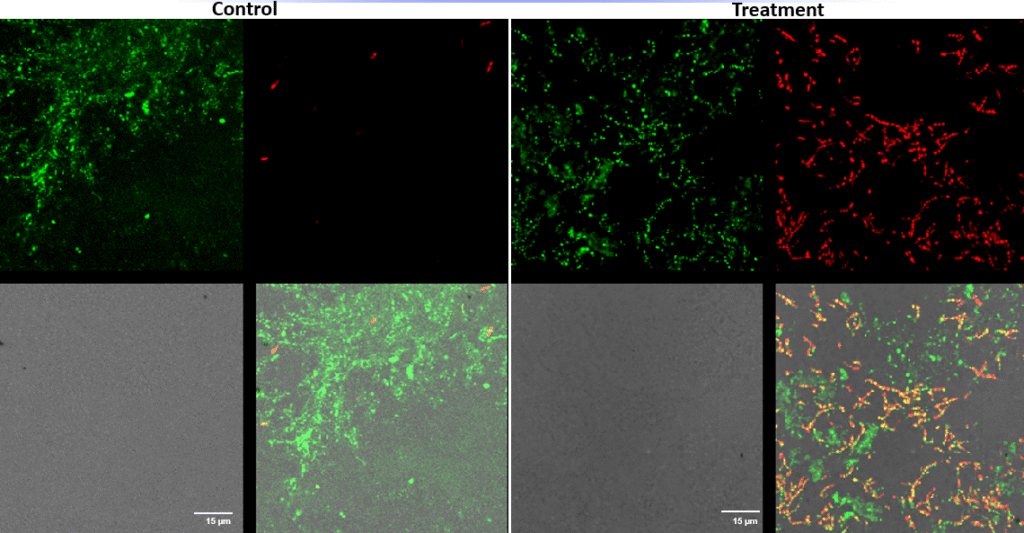
Pseudomonas aeruginosa – Superficial fluorescence microscopy. Microscopic imaging of Pseudomonas aeruginosa inoculated on the corneal surface and treated with UV-C for 15 seconds at a 10 mm distance. The images show clear bacterial membrane disruption and reduced viability, demonstrating the antimicrobial effectiveness of UV-C treatment on the superficial corneal layer.
Each image is divided into four quadrants: the upper left quadrant shows the green channel highlighting live bacteria; the upper right shows the red channel highlighting dead bacteria; the bottom right shows the merged green and red channels indicating live and dead bacteria; and the bottom left illustrates the bright field image. Bright green represents bacteria with intact membranes (live bacteria), red represents bacteria with compromised or dead membranes, and yellow represents bacteria with partially compromised membranes. Scale bar: 15 µm.
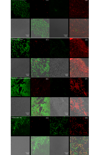
Panel with fluorescence microscope images of Pseudomonas aeruginosa (A & B), Streptococcus canis (C & D), Staphylococcus pseudintermedius (E & F), and a polymicrobial mix (G & H) inoculated superficially and treated with UV-C for 15 seconds.
Control samples are represented by images A, C, E, and G, and treated samples by images B, D, F, and H.
Each image is divided into four quadrants: the upper left quadrant shows the green channel highlighting live bacteria; the upper right shows the red channel highlighting dead bacteria; the bottom right shows the merged green and red channels highlighting live and dead bacteria; and the bottom left illustrates the bright field image. Bright green represents bacteria with intact membranes (live), red represents bacteria with compromised or dead membranes, and yellow represents bacteria with partially compromised membranes. Scale bar: 15 µm.
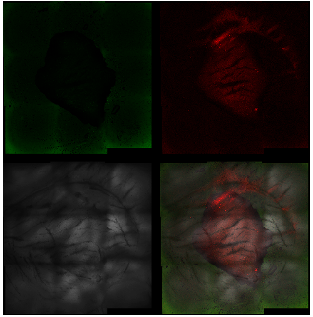
Fluorescence microscope images of the treated area with UV-C for 15 seconds (magnification 5×).
Each image is divided into four quadrants: the upper left quadrant shows the green channel highlighting live bacteria; the upper right shows the red channel highlighting dead bacteria; the bottom right shows the merged green and red channels indicating live and dead bacteria; and the bottom left illustrates the bright field image. Bright green represents bacteria with intact membranes (live bacteria), red represents bacteria with compromised or dead membranes, and yellow represents bacteria with partially compromised membranes.
It is noticeable that the central UV-C treated area contains predominantly dead bacteria, surrounded by live bacteria in the untreated area.
Hoerdemann M, Sahoo D.K., Allbaugh R.A., Kubai M.A. Ultraviolet C (UV-C) Light Therapy for Equine Ulcerative Keratomycosis – An In Vitro Study. Veterinary Ophthalmology. March 5, 2025; doi:10.1111/vop.70012.
Hoerdemann M., Sahoo D.K., Allbaugh R.A., Kubai M.A. Ultraviolet C (UV-C) Light Therapy for Equine Ulcerative Keratomycosis – An In Vitro Study. Veterinary Ophthalmology. March 5, 2025; doi:10.1111/vop.70012.
Our device is designed to support the treatment of corneal infections by providing an innovative adjunct therapy that complements standard medical approaches.
Yes. Safety is a key priority. The device has been developed with built-in protective measures and protocols, and its use is supported by peer-reviewed research in veterinary ophthalmology.
We encourage you to review the scientific papers referenced on this site, which provide detailed data and evidence supporting the clinical use of our technology.